lv m medical | lv wall thickness on echo lv m medical Normal values of left ventricular mass (LV M) and cardiac chamber sizes are prerequisites for the diagnosis of individuals with heart disease. LV M and cardiac chamber sizes may be recorded during cardiac computed tomography angiography (CCTA), and thus modality specific normal values are needed. The Canon LV-7292M is an LCD-based business projector with decent data and video image quality, and a relatively low brightness and price. Pros. Portable. Fairly good video quality for a data.
0 · lvh measurements in echo
1 · lvh echo criteria wall thickness
2 · lvh criteria on echo
3 · lv wall thickness on echo
4 · lv wall thickness echo measurement
5 · left ventricular wall thickness measurement
6 · how to calculate Lv mass
7 · causes of lvh on echo
If an educator needs to use the latest in 3D content in their classroom, the LV-8235 UST is ready with its 3D display system.The LV-8235 UST is Canon's first DLP projector, using a 0.65" single DLP chip and comes with Canon's commitment to ease of use and maintenance as well as our legendary optical prowess.
Left ventricular mass (LVM) has been shown to serve as a measure of target .
Left ventricular mass (LVM) is a well-established measure that can independently predict adverse cardiovascular events and premature death. 1-3 Population-based studies have revealed that increased LVM and left ventricular hypertrophy (LVH) as assessed by two-dimensional (2D) M-mode echocardiography measurements provide prognostic information . Left ventricular mass (LVM) has been shown to serve as a measure of target organ damage resulting from chronic exposure to several risk factors. Data on the association of midlife LVM with later cognitive performance are sparse.Our LV calculator allows you to painlessly evaluate the left ventricular mass, left ventricular mass index (LVMI for the heart), and the relative wall thickness (RWT). Read on and discover all the details of our LV mass calculator and its variables: Definitions of abnormal LV mass index; PWd normal range; and. IVSd in echo ️.
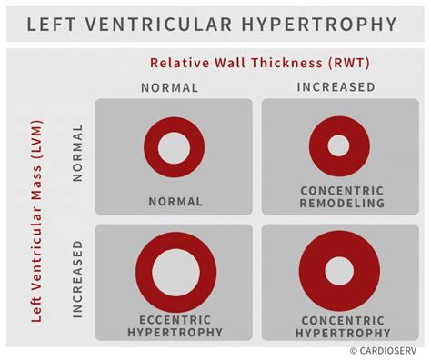
Normal values of left ventricular mass (LV M) and cardiac chamber sizes are prerequisites for the diagnosis of individuals with heart disease. LV M and cardiac chamber sizes may be recorded during cardiac computed tomography angiography (CCTA), and thus modality specific normal values are needed. Left ventricular mass (LVM) is a powerful predictor of cardiovascular risk. 1–4 LVM strongly relates to body size, indicating the need for appropriate normalization. 5,6 Because relations between body size and dimensions of organs are often nonlinear, allometric approaches are required, 5,6 in which LVM is divided by a body size variable . M-mode echocardiogram in left ventricular dysfunction. M-mode echocardiogram is commonly used to measure left ventricular dimensions and ejection fraction. Ejection fraction is indicative of the left ventricular systolic function.LVM is the acronym for Left Ventricular Mass. LV mass (LVM) is a vital prognostic measurement we obtain with echocardiography to manage hypertension. RWT is the acronym for Relative Wall Thickness and is an additional reference value that can help further classify the type of LVH.
The authors investigated 3 important areas related to the clinical use of left ventricular mass (LVM): accuracy of assessments by echocardiography and cardiac magnetic resonance (CMR), the ability to predict cardiovascular outcomes, and .LVMin in Medical commonly refers to Left Ventricular Mass, a critical measurement in assessing heart health and function, particularly in relation to hypertrophy and cardiovascular risk. This measurement is vital for diagnosing various cardiac conditions and monitoring treatment efficacy. The authors investigated 3 important areas related to the clinical use of left ventricular mass (LVM): accuracy of assessments by echocardiography and cardiac magnetic resonance (CMR), the ability to predict cardiovascular outcomes, and the comparative value of different indexing methods.
Left ventricular mass (LVM) is a well-established measure that can independently predict adverse cardiovascular events and premature death. 1-3 Population-based studies have revealed that increased LVM and left ventricular hypertrophy (LVH) as assessed by two-dimensional (2D) M-mode echocardiography measurements provide prognostic information . Left ventricular mass (LVM) has been shown to serve as a measure of target organ damage resulting from chronic exposure to several risk factors. Data on the association of midlife LVM with later cognitive performance are sparse.Our LV calculator allows you to painlessly evaluate the left ventricular mass, left ventricular mass index (LVMI for the heart), and the relative wall thickness (RWT). Read on and discover all the details of our LV mass calculator and its variables: Definitions of abnormal LV mass index; PWd normal range; and. IVSd in echo ️. Normal values of left ventricular mass (LV M) and cardiac chamber sizes are prerequisites for the diagnosis of individuals with heart disease. LV M and cardiac chamber sizes may be recorded during cardiac computed tomography angiography (CCTA), and thus modality specific normal values are needed.
Left ventricular mass (LVM) is a powerful predictor of cardiovascular risk. 1–4 LVM strongly relates to body size, indicating the need for appropriate normalization. 5,6 Because relations between body size and dimensions of organs are often nonlinear, allometric approaches are required, 5,6 in which LVM is divided by a body size variable .
M-mode echocardiogram in left ventricular dysfunction. M-mode echocardiogram is commonly used to measure left ventricular dimensions and ejection fraction. Ejection fraction is indicative of the left ventricular systolic function.LVM is the acronym for Left Ventricular Mass. LV mass (LVM) is a vital prognostic measurement we obtain with echocardiography to manage hypertension. RWT is the acronym for Relative Wall Thickness and is an additional reference value that can help further classify the type of LVH.The authors investigated 3 important areas related to the clinical use of left ventricular mass (LVM): accuracy of assessments by echocardiography and cardiac magnetic resonance (CMR), the ability to predict cardiovascular outcomes, and .
lvh measurements in echo
LVMin in Medical commonly refers to Left Ventricular Mass, a critical measurement in assessing heart health and function, particularly in relation to hypertrophy and cardiovascular risk. This measurement is vital for diagnosing various cardiac conditions and monitoring treatment efficacy.
lvh echo criteria wall thickness
ガリアーノ dior

туалетная вода dior

lvh criteria on echo
Product range . Support. Canon LV-X4. Download software, firmware and manuals and get access to troubleshooting resources for your projector. Software. Manuals. Firmware. FAQs & Help. Software (0) Software is an optional download that enables advanced functionality and helps you to get the most out of your product.
lv m medical|lv wall thickness on echo


