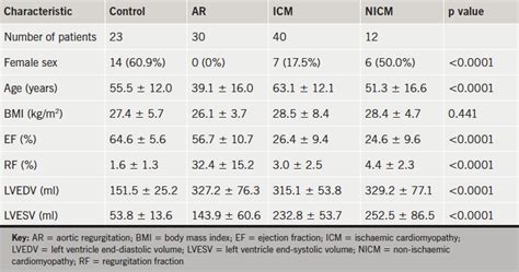
lv diameter echo | left internal dimension in systole lv diameter echo Normal 2D measurements: LV minor axis ≤ 2.8 cm/m 2, LV end-diastolic volume ≤ 82 ml/m 2, maximal LA antero-posterior diameter ≤ 2.8 cm/m 2, maximal LA volume ≤ 36 ml/m 2 (2;33;35). ∗∗ In the absence of other . Fairfield Inn & Suites by Marriott Saratoga Malta is located at 101 Saratoga Village Blvd. Fairfield Inn & Suites by Marriott Saratoga Malta has a variety of amenities that will .
0 · normal Lv size
1 · normal Lv end diastolic diameter
2 · normal Lv dimensions
3 · normal Lv diameter
4 · left internal dimension in systole
5 · echocardiography normal values pdf
6 · Lv cavity size
7 · 2d Lv pw abnormal
Parmi les événements importants de la Révolution française, citons la prise de la Bastille par le peuple, l'adoption de la Déclaration des droits de l'homme et du citoyen, le renversement de la monarchie et l'instauration de la République française, l'exécution du roi Louis XVI, le règne de la Terreur et les guerres révolutionnaires .
normal Lv size
Left and right ventricle. Visual assessment of systolic function. Visual assessment of regional wall motion (left ventricle) Recommended by American Society for Echocardiography (J Am Soc Echocardiogr 18:1440-1463, 2005). Left .(see below) and is derived from the LV end-diastolic volume (LVEDV) and LV end-systolic . The Left Ventricle. Example: Measurement end - Diastolic wall thickness (red) + .tricular [LV] size and ejection fraction [EF], left atrial [LA] volume), outcomes data are lacking .
Normal 2D measurements: LV minor axis ≤ 2.8 cm/m 2, LV end-diastolic volume ≤ 82 ml/m 2, maximal LA antero-posterior diameter ≤ 2.8 cm/m 2, maximal LA volume ≤ 36 ml/m 2 (2;33;35). ∗∗ In the absence of other .Assessment of left ventricular systolic function has a central role in the evaluation of cardiac disease: guiding management and predicting outcomes. •. Numerous echocardiographic techniques, including left ventricular ejection fraction, are .
rolex gmt master 2 von 2014 neupreis
Each of the following echo parameters are discussed and updated in turn: left ventricular linear .Echocardiographic assessment of the left ventricle (LV) begins with the measurement of linear . The first and most commonly used echocardiography method of LVM estimation is the linear method, which uses end-diastolic linear measurements of the interventricular septum (IVSd), LV inferolateral wall . Normal Values of Left Ventricular Mass by Two-Dimensional and Three .

Left and right ventricle. Visual assessment of systolic function. Visual assessment of regional wall motion (left ventricle) Recommended by American Society for Echocardiography (J Am Soc Echocardiogr 18:1440-1463, 2005). Left ventricular mass and geometry. Left ventricular dimension and volume. Left ventricular function (ejection fraction)(see below) and is derived from the LV end-diastolic volume (LVEDV) and LV end-systolic volume (LVESV). Global Longitudinal Strain is a new parameter to assess LV systolic function.
The Left Ventricle. Example: Measurement end - Diastolic wall thickness (red) + LV diameter (green) Each echocardiogram includes an evaluation of the LV dimensions, wall thicknesses and function. Good measurements are essential and may have implications for .tricular [LV] size and ejection fraction [EF], left atrial [LA] volume), outcomes data are lacking for many other parameters. Unfortunately, this approach also has limitations. Normal 2D measurements: LV minor axis ≤ 2.8 cm/m 2, LV end-diastolic volume ≤ 82 ml/m 2, maximal LA antero-posterior diameter ≤ 2.8 cm/m 2, maximal LA volume ≤ 36 ml/m 2 (2;33;35). ∗∗ In the absence of other etiologies of LV and LA dilatation and acute MR.Assessment of left ventricular systolic function has a central role in the evaluation of cardiac disease: guiding management and predicting outcomes. •. Numerous echocardiographic techniques, including left ventricular ejection fraction, are used in routine clinical practice to assess left ventricular systolic function. •.
Each of the following echo parameters are discussed and updated in turn: left ventricular linear dimensions and LV mass; left ventricular volumes; left ventricular ejection fraction; left atrial size; right heart parameters; aortic dimensions; and tissue Doppler imaging.
Echocardiographic assessment of the left ventricle (LV) begins with the measurement of linear dimensions that approximate its ellipsoid diameter. These linear dimensions have historically been measured at the basal level of the LV, which is not representative of its true diameter.
The first and most commonly used echocardiography method of LVM estimation is the linear method, which uses end-diastolic linear measurements of the interventricular septum (IVSd), LV inferolateral wall thickness, and LV internal diameter derived from 2D-guided M-mode or direct 2D echocardiography. Normal Values of Left Ventricular Mass by Two-Dimensional and Three-Dimensional Echocardiography: Results from the World Alliance Societies of Echocardiography Normal Values Study - Journal of the American Society of Echocardiography.Left and right ventricle. Visual assessment of systolic function. Visual assessment of regional wall motion (left ventricle) Recommended by American Society for Echocardiography (J Am Soc Echocardiogr 18:1440-1463, 2005). Left ventricular mass and geometry. Left ventricular dimension and volume. Left ventricular function (ejection fraction)
normal Lv end diastolic diameter
(see below) and is derived from the LV end-diastolic volume (LVEDV) and LV end-systolic volume (LVESV). Global Longitudinal Strain is a new parameter to assess LV systolic function. The Left Ventricle. Example: Measurement end - Diastolic wall thickness (red) + LV diameter (green) Each echocardiogram includes an evaluation of the LV dimensions, wall thicknesses and function. Good measurements are essential and may have implications for .
tricular [LV] size and ejection fraction [EF], left atrial [LA] volume), outcomes data are lacking for many other parameters. Unfortunately, this approach also has limitations. Normal 2D measurements: LV minor axis ≤ 2.8 cm/m 2, LV end-diastolic volume ≤ 82 ml/m 2, maximal LA antero-posterior diameter ≤ 2.8 cm/m 2, maximal LA volume ≤ 36 ml/m 2 (2;33;35). ∗∗ In the absence of other etiologies of LV and LA dilatation and acute MR.Assessment of left ventricular systolic function has a central role in the evaluation of cardiac disease: guiding management and predicting outcomes. •. Numerous echocardiographic techniques, including left ventricular ejection fraction, are used in routine clinical practice to assess left ventricular systolic function. •.Each of the following echo parameters are discussed and updated in turn: left ventricular linear dimensions and LV mass; left ventricular volumes; left ventricular ejection fraction; left atrial size; right heart parameters; aortic dimensions; and tissue Doppler imaging.
Echocardiographic assessment of the left ventricle (LV) begins with the measurement of linear dimensions that approximate its ellipsoid diameter. These linear dimensions have historically been measured at the basal level of the LV, which is not representative of its true diameter. The first and most commonly used echocardiography method of LVM estimation is the linear method, which uses end-diastolic linear measurements of the interventricular septum (IVSd), LV inferolateral wall thickness, and LV internal diameter derived from 2D-guided M-mode or direct 2D echocardiography.
normal Lv dimensions
normal Lv diameter

See 10 day weather forecast for Valletta. Foreca provides the most accurate forecast for any location.
lv diameter echo|left internal dimension in systole


